Wildlife Diseases
The DNR reminds Wisconsin that the virus that has caused highly pathogenic avian influenza (HPAI) in wild birds still exists in the state. Information on HPAI can be found on our Avian Influenza webpage.
Below is a list of diseases and other health issues commonly found in Wisconsin wildlife, or the DNR monitors for their occurrence. Click on a specific disease to learn more about its transmission, common signs observed in affected wildlife, management actions, associated public health concerns, and links to additional information.
You can help monitor the health of Wisconsin's wildlife by reporting your sightings of sick or dead wildlife to the DNR. To report a sick or dead wild bird or wild mammal, please use this survey form: Sick or Dead Wildlife Reporting Form. If you prefer, you can also contact the Wildlife Switchboard by emailing DNRWildlifeSwitchboard@wisconsin.gov or calling 608-267-0866. You will need to leave a message for the switchboard staff. In your message, please include the number of wild animals, the species (such as a raccoon or Canada goose), if they were sick or dead, the location where you saw them, and the county and your contact information. It is not necessary to report wildlife killed along roadways.
All Diseases Searchable Table
Amphibian & Reptile Diseases
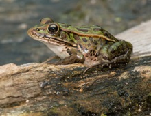
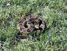
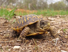
Image Examples of Amphibian & Reptile Species
Bird Diseases
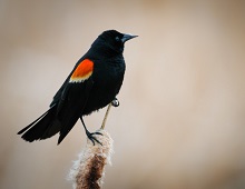
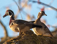
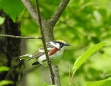
Image Examples of Bird Species
Bird Feeders: Keep birds healthy at feeders
If you find sick or dead songbirds (for example, finches, sparrows, siskins) at or near your bird feeder, it is possible they are getting sick from a disease that is easily transmitted when birds congregate in areas such as bird feeders, such as Salmonellosis or Trichomoniasis.
Please note that finding sick or dead songbirds at your feeder does not necessarily mean they got sick from anything you do if you follow the recommendations below. The birds may have arrived sick at your feeder and can now spread it to healthy birds arriving after them.
To break the cycle of sick birds infecting healthy birds, remove any bird feeders for 2 weeks. If no sick or dead birds are seen during the 2 weeks, follow the recommendations below before putting feeders back up.
- Wear gloves for personal protection.
- Wash feeders and birdbaths with soap and water.
- Disinfect feeders and birdbaths with a 10% bleach solution.
- Remove seed hulls and bird feces from under bird feeders and discard them in the trash.
- Move feeders occasionally to prevent the buildup of feces underneath the feeder.
- Add additional feeders to reduce overcrowding and contamination.
- Keep seeds and food dry.
- Change the water in birdbaths regularly.
Deer Diseases
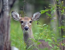
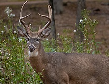
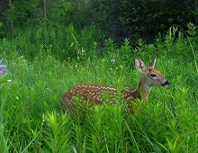
Image Examples of Deer Species
Other Mammal Diseases
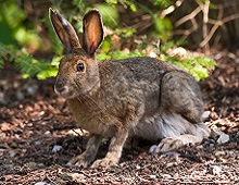
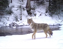
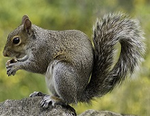
Image Examples of Other Mammals