Field Notes June 2025
A CLOSER LOOK AT THE DEATHS OF CWD-INFECTED DEER
Graphic Content Warning: This article shows up-close photos of dead deer that some readers may find upsetting.
Want to get notified when new newsletters come out? Sign up for the following topics on GovDelivery.
- Chronic Wasting Disease Updates - For emails about new project newsletters.
- Wisconsin Deer Research - For email updates on all deer projects being done by the Office of Applied Science.
For more Field Notes newsletters, please see the Field Notes Newsletter Index.
The Southwest Wisconsin CWD Study: How Specific Deer Illustrate Results
We examine the cases of three collared deer from the Southwest Wisconsin CWD, Deer and Predator Study, and explore how scientists can determine that their deaths were the result of CWD.
Deer #5010
We take a look at the case of deer #5010, a doe collared at 3.5 years old...
Deer #7235
Deer #7235 was collared at 8-9 months of age. After that, the study followed him for 20 months.
Deer #5006
Deer #5006 was collared as a 6.5-year-old doe. CWD was negative at collaring, and the study followed her life for almost two years.
The Southwest Wisconsin CWD Study: How Specific Deer Illustrate Results
By: Daniel Storm, PhD and Lead Scientist of the Southwest Wisconsin CWD, Deer and Predator Study
Earlier this year, we at the Wisconsin DNR announced the Southwest Wisconsin CWD, Deer and Predator Study's primary findings at the January Natural Resources Board meeting. This study conclusively demonstrated that CWD reduces deer survival, and it corroborated what studies from other states have found, which is also occurring in Wisconsin. Of course, CWD can reduce survival by killing deer outright, aka "dying of CWD," but it can also do so by making them more vulnerable to hunters, cars, coyotes and so on. In this article, we won't cover all the various ways deer can die. Instead, we'll dive into the gory details of deer that died directly from CWD. And I do mean gory. Talking about disease and mortality and demonstrating all the evidence necessary to conclude a deer died of a specific cause unavoidably involves photos and descriptions of dead deer. So, warning: It ain't pretty! However, if you've ever wondered to yourself, "Do deer actually die from CWD?," "How do deer die of CWD?", or "If so many deer are that sick, why don't I see them everywhere?," then this article is for you.
To explore this topic, I will show and discuss individual deer from the study. We'll start with what we knew about them at capture, then go into how long they lived, what our investigations and necropsy reports revealed, and any other interesting notes about them.
First, a quick review on how we collect this information:
- Catch and Collar the Deer – This includes an antemortem "before death" test for CWD, which we obtained by taking a small piece of tissue from the rectum (yes, you read that right) and sending it to the Wisconsin Veterinary Diagnostic Laboratory for CWD testing. We then placed GPS collars on the deer, providing high-quality information about their movements. Finally, we weighed the deer and recorded a simple body condition index, which indicated how fat or lean each deer was.
- Investigate Mortalities – A feature of GPS collars is that when the collar becomes motionless (indicating deer death), we get text and email alerts, which allows us to find the deer as soon as possible. If the deer were at least mostly intact, we took the carcass to the UW-Madison School of Veterinary Medicine, where a veterinary pathologist would conduct a complete necropsy (animal autopsy). If the carcass were too heavily consumed or rotten, we would still do as extensive a field necropsy as possible and look for evidence such as infections, bite marks, broken bones or anything else that might clue us into what happened with the deer. We always conducted what amounted to a crime-scene investigation of the surrounding area, looking for any information on how the animal died or what might have contributed to its death.
For this article, we'll focus on deer that had a necropsy report from UW-Madison because we have the most information about these deer. Okay, let's get started.
Deer #5010
The first one is deer #5010, a doe we collared on Dec. 28, 2017, when she was about 3.5 years of age. Interestingly, at capture, she only weighed 118 pounds, much lower than the average capture weight for adult does, which is 144 pounds. Her body condition score was 0 out of 10. This basically means that the technicians could feel her backbone easily, and that she lacked body fat in this location. This is a quick-and-dirty, but effective, way to assess body condition in deer. A low body condition score is not necessarily uncommon or indicative of imminent starvation, but it is unusual to see it in a prime-aged deer so early in winter. So this doe was not in great shape, but as we'll see, she had much further to fall. The last thing I'll note is that she in no other way appeared outwardly sick to the technicians at capture – she was not stumbling or drooling, and her head and ears were not drooping.
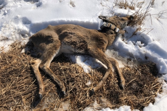
Fast forward almost seven weeks to Feb. 14, 2018; #5010 was dead and looked much worse for the wear. This photo shows just how emaciated she was.
Note the hips sticking out and the sunken shoulder and rump. Here is a quote from the necropsy report:
"The animal is in poor overall body condition with 0 mm of subcutaneous fat at the xiphoid and rump. There is additionally inapparent perirenal, heart, omental and retrobulbar fat."
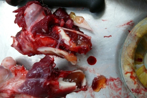
This is all a technical way of saying the deer had no fat right under the skin (subcutaneous) and anywhere within the body cavity where they would typically expect to find fat. Further, this photo shows the bone marrow, which looks like red jelly. Usually, this would be white and chalky, full of fat. This is the last reserve of fat in the body, and what biologists check to confirm starvation – if a deer had red jelly bone marrow, it was starving. She weighed a scant 78 pounds at death, which is a 40-pound loss in just 7 weeks.
Some interesting notes:
- She had one fetus. She likely became pregnant between 2.5 and 3.5 months before her death, so her disease state at just a handful of months before her death was not so severe as to prevent her from getting pregnant. This is another piece of evidence that CWD affects deer briefly, relative to the entire course of infection, which is thought to take 18-24 months.
- She did have food in her digestive system, which is somewhat interesting, considering that she starved. However, we don't know if her food intake was simply inadequate or if she could not digest her food properly.
- "…coat is unkempt with numerous live lice…" One of the findings from our 2022 publication, Cause of death, pathology, and chronic wasting disease status of white-tailed deer (Odocoileus virginianus) mortalities in Wisconsin, USA, was that deer with CWD were more likely to have parasites like lice and ticks. This could happen because CWD-positive deer are not grooming themselves as much as normal or receive less grooming from other deer. It could also be because their immune systems are less able to fend off parasites.
- One portion of the right lung had an abscess, a localized infection, but they considered this to have "...limited overall health implications."
- They found some lungworms, but this infection was considered mild.
Additionally, the following quote, also from the necropsy report, speaks to the deer having CWD:
"Emaciation and histologic lesions of neuronal vacuolation are consistent with Chronic Wasting Disease, and CWD
immunohistochemistry was positive in both the brainstem and the retropharyngeal lymph node."
"Neuronal vacuolation" is the technical term for the holes in the brain that are characteristic of CWD. "Immunohistochemistry" is the type of CWD test used, which is considered the gold standard. The bottom line with all of this, is that the deer 1.) starved, and 2.) had CWD. This is a classic end-stage chronic wasting disease death.
Deer #7235
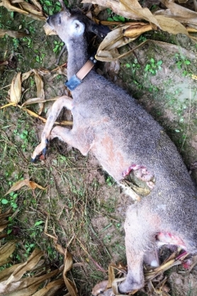
Next up, deer #7235 – and even by the standards of dead deer, this is a gross one. We captured him on Feb. 11, 2017, when he was 8-9 months of age. He had a body condition score of 1/10 and weighed 86 pounds at capture, which is pretty close to average for a buck this age. At the time, CWD was not detected in his biopsy sample. On Oct. 26, 2017, he dispersed about 3 miles from his initial home range. We received a mortality alert almost 20 months after capture, on Sept. 30, 2018. One of our field technicians found his carcass a few rows into a cornfield. As can be seen in the photo below, he'd been scavenged a bit and there were coyote tracks in the mud, indicating the likely culprit. So, how can we tell he was scavenged post-death and not killed by the coyotes? There are a few ways. A depredation event usually involves struggle, which would have been apparent in the form of torn-up ground, lots of knocked-over corn stalks, hair and bits of flesh strewn about. None of that is present. Instead, it appears as though the deer lay down and died, and then the coyotes came in and started feeding. Lastly, there were no bite marks on the neck, head or any place other than where the deer was eaten, nor was there any evidence of bleeding anywhere on the body.
Despite the scavenging, we could still get a necropsy report from the UW vet school. He tested positive for CWD and exhibited "...diffuse moderate spongiosis with intraneuronal vacuoles and gliosis…". "Spongiosis'" means essentially the same thing as vacuolation, the holes in the brain, and "gliosis" refers to the formation of scar tissue in the brain in response to damage. Like all end-stage CWD-infected deer, he was also emaciated.
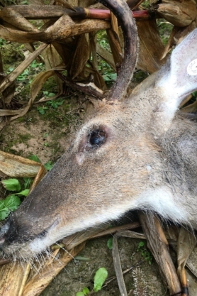
"The body is in poor nutritional condition with inapparent visceral and subcutaneous fat and moderate muscle wasting. The eyes are sunken and the skull bone prominent due to fat and muscle atrophy."
Note how this deer became emaciated at a time when food was everywhere, meaning CWD, not lack of food, caused this starvation. That's not all that was going wrong with this buck. However, he also suffered from "Severe bilateral cranioventral fibrinosuppurative bronchopneumonia." This means that both the front and the bottom portions of both lungs were infected and filled with clots and pus. Like I said, it ain't pretty. Unlike #5010, pneumonia significantly contributed to this deer's death. It's worthwhile to pause here and consider why these end-stage deer are getting pneumonia. It is generally thought to result from inhaling saliva, water and food, which they seem to do more often in the later stages of CWD because they salivate more and have trouble swallowing. These foreign substances introduce lots of bacteria into the lungs, which sets off the pneumonia.
A few additional notes of interest from the necropsy:
- His antlers were notably small for a 2-year-old buck. At the time of his death which, on Sept. 30, he was shedding velvet, which is on the late side to still have antler velvet. This looks like an instance when the timing of antler development lined up with the end-stage of CWD, leading to poor antler growth. Since this buck was starving, he lacked the energy to put towards antler growth.
- Like #5010, this deer also had food in his stomach.
One last note on this buck: his range in the previous 2 weeks of his life was about 80 acres (an eighth of a square mile). Over this same timeframe, bucks without CWD had an average range of 345 acres (a little over half a square mile). It appears that his being in the late stages of CWD caused him to move significantly less, which makes a lot of sense; he was starving, so he had less energy, but is this drop in range size typical? In what other ways does CWD change deer movement behavior? We are currently completing an analysis that answers these questions and will have an article in the near future that lays this all out. Stay tuned!
Deer #5006
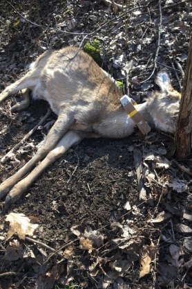
Lastly, we'll look at deer #5006. We first captured #5006 as a 6.5-year-old doe on Jan. 9, 2017. At that time, she tested CWD-negative, weighed 165 pounds and had a body condition score of 5/10. We then captured her again about 2 years later, on Feb. 4, 2019. She hadn't changed much in terms of body weight and condition, weighing 164 pounds with a 7/10 body condition score. This time she tested CWD-positive, but by all outward appearances was still a big, healthy doe. Fifty days later, however, she was dead. She looked like this when the technicians found her lying on her side, untouched.
At necropsy, her carcass weighed 90 pounds, which is a loss of 74 pounds (45% of her body weight) in just 49 days. Unsurprisingly, she had "scant to inapparent subcutaneous and visceral adipose stores and moderate muscle wasting." Note: "adipose" means fat. She also had pneumonia, specifically "Marked multifocal to extensive necrosuppurative bronchopneumonia,, which basically means pneumonia was found throughout her lungs and involved dead lung tissue and pus. Lastly, they found the holes in the brain associated with CWD "spongiform encephalopathy with intracytoplasmic neuronal vacuoles" and her post-mortem CWD test was positive. Deer #5006 presents a classic case of end-stage CWD.
These deer are just three examples, but I think they illustrate just how devastating CWD is on an individual level. They show that end-stage CWD always involves starvation and oftentimes other infections, like pneumonia. They also demonstrate just how fast the end-stage of CWD is, considering that it takes 18 months or more for deer to reach that end-stage once they've been infected. The speed of this decline explains why, despite high prevalence numbers, people might not be seeing many "sick" deer on the landscape.


